

How much does a human brain weight?

What is the human brain made of?
The human mind is the focal organ of the human sensory system, and the spinal line makes up the focal sensory device. The thoughts accommodate the frontal cortex, the brainstem, and the cerebellum. It controls the greater part of the sporting activities of the body, preparing, incorporating, and planning the statistics it receives from the receptors and deciding on picks concerning the pointers shipped off the rest of the body. The cerebrum is contained in, and ensured with the aid of, the skull bones of the top.
The frontal cortex, the largest piece of the human cerebrum, contains two cerebral halves of the globe. Every half of the globe has an inner center produced from white matter, and an external floor – the cerebral cortex – produced from dim matter. The cortex has an outside layer, the neocortex, and an inward allocortex. The neocortex is comprised of six neuronal layers, whilst the allocortex has 3 or four. Every 1/2 of the globe is typically isolated into 4 flaps – the front going through, worldly, parietal, and occipital projections. The front dealing with projection is related with chief capacities together with poise, arranging, thinking, and particular idea, at the same time as the occipital flap is devoted to imagination and prescient. Inside each projection, cortical regions are related with express capacities, just like the tactile, engine, and association areas. Albeit the left and right aspects of the equator are comprehensively similar suit and capability, a few capacities are associated with one side, like language inside the left and visual-spatial potential morally justified. The halves of the globe are related by using commissural nerve plots, the largest being the corpus callosum.
The frontal cortex is associated via the brainstem to the spinal string. The brainstem comprises the midbrain, the pons, and the medulla oblongata. The cerebellum is associated with the brainstem by units of plots. Inside the frontal cortex is the ventricular framework, comprising of 4 interconnected ventricles in which cerebrospinal liquid is created and turned around. Under the cerebral cortex is a few massive buildings, including the thalamus, the epithalamus, the pineal organ, the nerve middle, the pituitary organ, and the subthalamus; the limbic designs, which includes the amygdala and the hippocampus; the claustrum, the one of a kind cores of the basal ganglia; the basal forebrain systems, and the 3 circumventricular organs. The cells of the cerebrum contain neurons and consistent glial cells. There are an extra 86 billion neurons in the cerebrum and a quite plenty equal number of different cells. Mind movement is made doable by using the interconnections of neurons and their arrival of synapses in mild nerve riding forces. Neurons companion with shape neural pathways, neural circuits, and problematic agency frameworks. The complete hardware is driven by using the interplay of neurotransmission.
The cerebrum is secured via the cranium, suspended in cerebrospinal liquid, and restrained from the circulation device by means of the blood–mind quandary. In any case, the mind is as yet defenseless to harm, sickness, and disease. Harm can be delivered approximately through damage, or a deficiency of blood delivery called a stroke. The mind is helpless to degenerative problems, like Parkinson's illness, dementias which include Alzheimer's illness, and numerous sclerosis. Mental conditions, such as schizophrenia and clinical disappointment, are believed to be associated with cerebrum dysfunctions. The thoughts can likewise be the website of tumors, both benevolent and dangerous; those generally start from extraordinary destinations in the frame.
The investigation of the lifestyles systems of the mind is neuroanatomy, whilst the research of its capability is neuroscience. Various strategies are utilized to do not forget the cerebrum. Examples from distinctive creatures, which might be inspected infinitesimally, have commonly given a lot of facts. Clinical imaging improvements, for example, practical neuroimaging, and electroencephalography (EEG) chronicles are substantial in examining the cerebrum. The medical history of individuals with mind harm has given knowledge into the ability of every piece of the cerebrum. Cerebrum studies have developed over the lengthy haul, with philosophical, check, and hypothetical tiers. A bobbing-up degree might be to reenact cerebrum movement.
In tradition, the way of taking into account the psyche has for pretty a long term endeavored to solve the difficulty of the concept of consciousness and the brain frame trouble. The pseudoscience of phrenology endeavored to limit man or woman credit to locales of the cortex within the nineteenth century. In sci-fi, cerebrum transfers are envisioned in tales, for instance, the 1942 Donovan's Brain.
Structure

The adult human mind weighs on average about 1.2–1.4 kg (2.6–3.1 lb) which is set 2% of the full body weight,[4][5] with a volume of around 1260 cm3 in men and 1130 cm3 in girls.[6] There is a giant character variation,[6] with the same old reference variety for guys being 1, a hundred and eighty–1,620 g (2.60–three.57 lb)and for girls 1,030–1, four hundred g (2.27–3.09 lb).
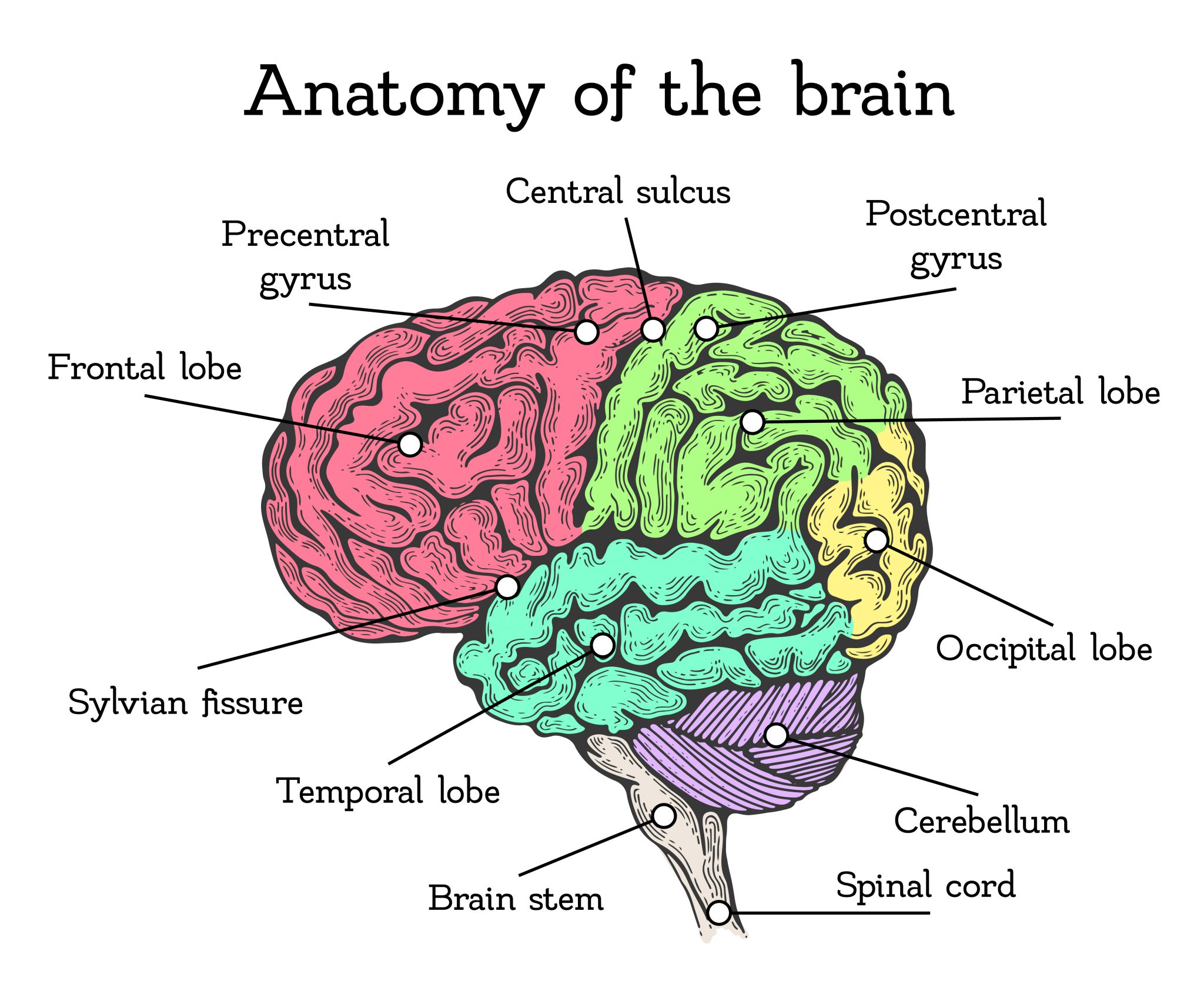
The cerebrum, consisting of the cerebral hemispheres, bureaucracy the largest part of the mind and overlies the alternative brain structures. The outer place of the hemispheres, the cerebral cortex, is gray rely upon, including cortical layers of neurons. Each hemisphere is divided into 4 foremost lobes – the frontal lobe, parietal lobe, temporal lobe, and occipital lobe. Three different lobes are included by means of a few resources which are a primary lobe, a limbic lobe, and an insular lobe. The relevant lobe accommodates the precentral gyrus and the postcentral gyrus and is covered since it bureaucracy a distinct purposeful role.
The brainstem, corresponding to a stalk, attaches to and leaves the cerebrum at the beginning of the midbrain region. The brainstem includes the midbrain, the pons, and the medulla oblongata. Behind the brainstem is the cerebellum.
The cerebrum, brainstem, cerebellum, and spinal cord are included by way of 3 membranes referred to as meninges. The membranes are the tough dura mater; the middle arachnoid mater and the extra sensitive inner pia mater. Between the arachnoid mater and the pia mater are the subarachnoid area and subarachnoid cisterns, which incorporate the cerebrospinal fluid. The outermost membrane of the cerebral cortex is the basement membrane of the pia mater called the glia limitans and is an important part of the blood–mind barrier. The dwelling brain may be very smooth, having a gel-like consistency similar to smooth tofu. The cortical layers of neurons constitute plenty of the cerebral gray relies upon, even as the deeper subcortical areas of myelinated axons, make up the white count number. The white count number of the brain makes up approximately half of the total mind quantity.
Cerebrum

The cerebrum is the biggest part of the brain and is divided into almost symmetrical left and right hemispheres by way of a deep groove, the longitudinal fissure. Asymmetry among the lobes is cited as a petalia. The hemispheres are related by using five commissures that span the longitudinal fissure, the biggest of these is the corpus callosum. Each hemisphere is conventionally divided into four important lobes; the frontal lobe, parietal lobe, temporal lobe, and occipital lobe, named consistent with the cranium bones that overlie them.[10] Each lobe is related to one or two specialized functions although there may be some useful overlap among them. The floor of the brain is folded into ridges (gyri) and grooves (sulci), a lot of that is named, typically according to their role, together with the frontal gyrus of the frontal lobe or the primary sulcus keeping apart the central regions of the hemispheres. There are many small versions in the secondary and tertiary folds.
The outer part of the cerebrum is the cerebral cortex, made of gray matter organized in layers. It is two to four millimeters (zero.079 to 0.157 in) thick and deeply folded to give a convoluted look. Beneath the cortex is the cerebral white count number. The largest part of the cerebral cortex is the neocortex, which has six neuronal layers. The rest of the cortex is of the allocortex, which has 3 or 4 layers.
The cortex is mapped through divisions into about fifty unique functional regions referred to as Brodmann's areas. These areas are quite extraordinary while visible beneath a microscope. The cortex is divided into foremost purposeful regions – a motor cortex and a sensory cortex. The primary motor cortex, which sends axons all the way down to motor neurons in the brainstem and spinal wire, occupies the rear part of the frontal lobe, immediately in front of the somatosensory place. The number one sensory area acquires signals from the sensory nerves and tracts by way of relay nuclei in the thalamus. Primary sensory areas include the visual cortex of the occipital lobe, the auditory cortex in components of the temporal lobe and insular cortex, and the somatosensory cortex inside the parietal lobe. The remaining components of the cortex, are known as the affiliation areas. These regions acquire input from the sensory areas and decrease components of the mind and are involved within the complicated cognitive strategies of belief, thought, and selection-making. The predominant features of the frontal lobe are to control attention, abstract thinking, behavior, trouble fixing tasks, and physical reactions and character. The occipital lobe is the smallest lobe; its primary features are visual reception, visual-spatial processing, motion, and shade reputation. There is a smaller occipital lobule inside the lobe known as the cuneus. The temporal lobe controls auditory and visual reminiscences, language, and some listening to and speech.
The cerebrum incorporates the ventricles in which the cerebrospinal fluid is produced and circulated. Below the corpus callosum is the septum pellucidum, a membrane that separates the lateral ventricles. Beneath the lateral ventricles is the thalamus and to the front and underneath this is the hypothalamus. The hypothalamus leads directly to the pituitary gland. At the lower back of the thalamus is the brainstem.
The basal ganglia, additionally called basal nuclei, are a set of structures deep inside the hemispheres concerned with behavior and emotion regulation. The largest element is the striatum, others are the globus pallidus, the substantia nigra, and the subthalamic nucleus. The striatum is divided into ventral striatum, and dorsal striatum, subdivisions which might be based totally upon function and connections. The ventral striatum consists of the nucleus accumbens and the olfactory tubercle while the dorsal striatum consists of the caudate nucleus and the putamen. The putamen and the globus pallidus lie separated from the lateral ventricles and thalamus by the inner tablet, while the caudate nucleus stretches around and abuts the lateral ventricles on their outer facets. At the private part of the lateral sulcus among the insular cortex and the striatum is a thin neuronal sheet known as the claustrum.
Below and in front of the striatum are some basal forebrain structures. These consist of the nucleus basalis, diagonal band of Broca, substantia innominata, and the medial septal nucleus. These systems are essential in producing the neurotransmitter, acetylcholine, which is then allotted broadly throughout the mind. The basal forebrain, especially the nucleus basalis, is taken into consideration to be the fundamental cholinergic output of the relevant frightened device to the striatum and neocortex.
Cerebellum
The cerebellum is split into an anterior lobe, a posterior lobe, and the flocculonodular lobe. The anterior and posterior lobes are connected within the middle with the aid of the vermis. Compared to the cerebral cortex, the cerebellum has a much thinner outer cortex that is narrowly furrowed into numerous curved transverse fissures. Viewed from below between the two lobes is the 1/3 lobe the flocculonodular lobe. The cerebellum rests in the back of the cranial hollow space, mendacity beneath the occipital lobes, and is separated from these via the cerebellar tentorium, a sheet of fiber.
It is connected to the midbrain of the brainstem by means of the superior cerebellar peduncles, to the pons by using the center cerebellar peduncles, and to the medulla by using the inferior cerebellar peduncles. The cerebellum consists of an inner medulla of white matter and an outer cortex of richly folded grey count. The cerebellum's anterior and posterior lobes seem to play a position inside the coordination and smoothing of complex motor moves, and the flocculonodular lobe within the maintenance of stability despite the fact that debate exists as to its cognitive, behavioral and motor features.
Brainstem
The brainstem lies under the cerebrum and consists of the midbrain, pons, and medulla. It lies within the lower back part of the skull, resting on the part of the base referred to as the clivus, and ends at the foramen magnum, a big beginning within the occipital bone. The brainstem maintains under this as the spinal wire, included by the vertebral column.
Ten of the twelve pairs of cranial nerves[a] emerge directly from the brainstem. The brainstem also consists of many cranial nerve nuclei and nuclei of peripheral nerves, as well as nuclei involved within the regulation of many vital techniques together with respiration, control of eye actions, and balance.[40][39] The reticular formation, a network of nuclei of sick-described formation, is a gift inside and alongside the period of the brainstem.[39] Many nerve tracts, which transmit data to and from the cerebral cortex to the rest of the body, pass through the brainstem.
Microanatomy

The human brain is commonly composed of neurons, glial cells, neural stem cells, and blood vessels. Types of neuron encompass interneurons, pyramidal cells consisting of Betz cells, motor neurons (upper and lower motor neurons), and cerebellar Purkinje cells. Betz cells are the largest cells (via size of mobile frame) within the nervous system. The adult human mind is estimated to incorporate 86±8 billion neurons, with a kind of same range (eighty five±10 billion) of non-neuronal cells. Out of these neurons, 16 billion (19%) are placed inside the cerebral cortex, and 69 billion (80%) are within the cerebellum.
Types of glial cell are astrocytes (which includes Bergmann glia), oligodendrocytes, ependymal cells (together with tanycytes), radial glial cells, microglia, and a subtype of oligodendrocyte progenitor cells. Astrocytes are the most important of the glial cells. They are stellate cells with many processes radiating from their cell our bodies. Some of these tactics cease as perivascular cease-toes on capillary walls.The glia limitans of the cortex is made of astrocyte foot techniques that serve in component to include the cells of the mind.
Mast cells are white blood cells that have interaction in the neuroimmune system in the brain. Mast cells in the primary frightened device are present in some of systems such as the meninges; they mediate neuroimmune responses in inflammatory conditions and assist to keep the blood–mind barrier, specially in mind regions wherein the barrier is absent. Mast cells serve the equal wellknown functions inside the body and imperative nervous machine, which include effecting or regulating allergic responses, innate and adaptive immunity, autoimmunity, and inflammation. Mast cells serve as the main effector mobile thru which pathogens can have an effect on the biochemical signaling that takes area between the gastrointestinal tract and the crucial apprehensive machine.
Some 400 genes are shown to be brain-particular. In all neurons, ELAVL3 is expressed, and in pyramidal neurons, NRGN and REEP2 also are expressed. GAD1 – crucial for the biosynthesis of the neurotransmitter GABA – is expressed in interneurons. Proteins expressed in glial cells consist of astrocyte markers GFAP and S100B while myelin primary protein and the transcription component OLIG2 are expressed in oligodendrocytes
Cerebrospinal fluid
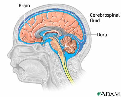
Cerebrospinal fluid is a clear, colourless transcellular fluid that circulates across the brain inside the subarachnoid space, in the ventricular device, and in the relevant canal of the spinal twine. It additionally fills some gaps in the subarachnoid area, called subarachnoid cisterns. The four ventricles, two lateral, a third, and a fourth ventricle, all include a choroid plexus that produces cerebrospinal fluid.The 0.33 ventricle lies in the midline and is connected to the lateral ventricles. A single duct, the cerebral aqueduct among the pons and the cerebellum, connects the 0.33 ventricle to the fourth ventricle. Three separate openings, the middle and two lateral apertures, drain the cerebrospinal fluid from the fourth ventricle to the cisterna magna one of the major cisterns. From right here, cerebrospinal fluid circulates across the mind and spinal twine in the subarachnoid space, among the arachnoid mater and pia mater. At any individual time, there may be approximately 150mL of cerebrospinal fluid – maximum within the subarachnoid space. It is continuously being regenerated and absorbed, and is replaced approximately as soon as every 5–6 hours.
A glymphatic gadget has been described as the lymphatic drainage system of the brain. The mind-huge glymphatic pathway includes drainage routes from the cerebrospinal fluid, and from the meningeal lymphatic vessels which might be related to the dural sinuses, and run alongside the cerebral blood vessels.[55][56] The pathway drains interstitial fluid from the tissue of the mind.
Blood deliver

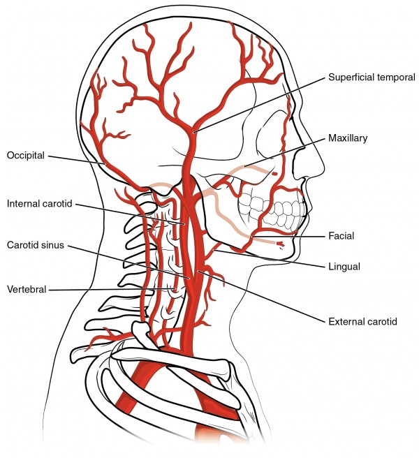
The internal carotid arteries supply oxygenated blood to the the front of the brain and the vertebral arteries deliver blood to the back of the mind. These circulations join together inside the circle of Willis, a ring of linked arteries that lies inside the interpeduncular cistern between the midbrain and pons.
The internal carotid arteries are branches of the commonplace carotid arteries. They input the skull through the carotid canal, travel through the cavernous sinus and input the subarachnoid space.They then enter the circle of Willis, with branches, the anterior cerebral arteries emerging. These branches journey ahead after which upward along the longitudinal fissure, and deliver the the front and midline elements of the brain. One or more small anterior communicating arteries be part of the 2 anterior cerebral arteries shortly after they come to be branches. The inner carotid arteries continue ahead because the center cerebral arteries. They journey sideways along the sphenoid bone of the eye socket, then upwards thru the insula cortex, wherein very last branches get up. The center cerebral arteries ship branches along their length.
The vertebral arteries turn out to be branches of the left and proper subclavian arteries. They tour upward thru transverse foramina which might be areas in the cervical vertebrae. Each aspect enters the cranial hollow space via the foramen magnum along the corresponding facet of the medulla. They supply off one of the three cerebellar branches. The vertebral arteries be a part of in front of the center part of the medulla to form the larger basilar artery, which sends more than one branches to supply the medulla and pons, and the two different anterior and superior cerebellar branches. Finally, the basilar artery divides into posterior cerebral arteries. These journey outwards, across the advanced cerebellar peduncles, and alongside the top of the cerebellar tentorium, where it sends branches to supply the temporal and occipital lobes.Each posterior cerebral artery sends a small posterior speaking artery to join with the internal carotid arteries.
Blood drainage
Cerebral veins drain deoxygenated blood from the brain. The mind has two predominant networks of veins: an exterior or superficial community, on the floor of the cerebrum that has three branches, and an indoors network. These two networks speak via anastomosing (becoming a member of) veins.The veins of the mind drain into large cavities of the dural venous sinuses normally located among the dura mater and the overlaying of the skull.Blood from the cerebellum and midbrain drains into the exquisite cerebral vein. Blood from the medulla and pons of the brainstem have a variable sample of drainage, either into the spinal veins or into adjoining cerebral veins.
The blood within the deep a part of the brain drains, thru a venous plexus into the cavernous sinus at the the front, and the advanced and inferior petrosal sinuses at the sides, and the inferior sagittal sinus on the back. Blood drains from the outer mind into the huge advanced sagittal sinus, which rests within the midline on pinnacle of the brain. Blood from right here joins with blood from the directly sinus at the confluence of sinuses.
Blood from here drains into the left and right transverse sinuses. These then drain into the sigmoid sinuses, which acquire blood from the cavernous sinus and superior and inferior petrosal sinuses. The sigmoid drains into the big internal jugular veins.
The blood–mind barrier

The large arteries in the course of the brain deliver blood to smaller capillaries. These smallest of blood vessels within the brain, are coated with cells joined through tight junctions and so fluids do not seep in or leak out to the equal diploma as they do in other capillaries; this creates the blood–brain barrier. Pericytes play a main role within the formation of the tight junctions.The barrier is much less permeable to larger molecules, but remains permeable to water, carbon dioxide, oxygen, and maximum fat-soluble materials (including anaesthetics and alcohol). The blood-mind barrier is not gift inside the circumventricular organs—which might be structures within the mind which can want to reply to modifications in body fluids—consisting of the pineal gland, location postrema, and a few regions of the hypothalamus. There is a similar blood–cerebrospinal fluid barrier, which serves the identical cause as the blood–mind barrier, however allows the transport of various substances into the mind because of the wonderful structural traits between the two barrier structures.
Development

At the start of the 0.33 week of improvement, the embryonic ectoderm forms a thickened strip called the neural plate. By the fourth week of development the neural plate has widened to offer a wide cephalic give up, a much less wide center element and a narrow caudal quit. These swellings are called the number one brain vesicles and represent the beginnings of the forebrain (prosencephalon), midbrain (mesencephalon), and hindbrain (rhombencephalon).
Neural crest cells (derived from the ectoderm) populate the lateral edges of the plate on the neural folds. In the fourth week—at some stage in the neurulation stage—the neural folds near shape the neural tube, bringing collectively the neural crest cells on the neural crest.The neural crest runs the period of the tube with cranial neural crest cells at the cephalic stop and caudal neural crest cells at the tail. Cells detach from the crest and migrate in a craniocaudal (head to tail) wave inside the tube. Cells on the cephalic cease supply rise to the mind, and cells at the caudal stop give rise to the spinal twine.
The tube flexes because it grows, forming the crescent-shaped cerebral hemispheres at the top. The cerebral hemispheres first appear on day 32. Early in the fourth week, the cephalic element bends sharply ahead in a cephalic flexure. This flexed component will become the forebrain (prosencephalon); the adjoining curving element will become the midbrain (mesencephalon) and the element caudal to the flexure turns into the hindbrain (rhombencephalon). These areas are shaped as swellings referred to as the three number one mind vesicles. In the 5th week of development 5 secondary mind vesicles have fashioned. The forebrain separates into two vesicles – an anterior telencephalon and a posterior diencephalon. The telencephalon gives rise to the cerebral cortex, basal ganglia, and related structures. The diencephalon gives rise to the thalamus and hypothalamus. The hindbrain additionally splits into two regions – the metencephalon and the myelencephalon. The metencephalon offers upward thrust to the cerebellum and pons. The myelencephalon offers upward thrust to the medulla oblongata. Also at some point of the fifth week, the brain divides into repeating segments referred to as neuromeres. In the hindbrain those are called rhombomeres.
A function of the mind is the cortical folding called gyrification. For simply over 5 months of prenatal improvement the cortex is easy. By the gestational age of 24 weeks, the wrinkled morphology displaying the fissures that start to mark out the lobes of the brain is evident. Why the cortex wrinkles and folds isn't nicely-understood, however gyrification has been linked to intelligence and neurological issues, and some of gyrification theories were proposed. These theories consist of those based totally on mechanical buckling, axonal tension, and differential tangential expansion. What is clear is that gyrification is not a random technique, however alternatively a complicated developmentally predetermined process which generates styles of folds which are regular among individuals and most species.
The first groove to appear within the fourth month is the lateral cerebral fossa. The expanding caudal cease of the hemisphere has to curl over in a ahead route to match into the restricted space. This covers the fossa and turns it into a much deeper ridge called the lateral sulcus and this marks out the temporal lobe. By the sixth month different sulci have formed that demarcate the frontal, parietal, and occipital lobes. A gene gift inside the human genome (ARHGAP11B) may play a primary position in gyrification and encephalisation.
Brain size

The length of the brain and someone's intelligence aren't strongly associated. Studies have a tendency to suggest small to moderate correlations (averaging round zero.Three to zero.Four) among brain quantity and IQ. The most constant associations are observed in the frontal, temporal, and parietal lobes, the hippocampi, and the cerebellum, but those handiest account for a notably small quantity of variance in IQ, which itself has handiest a partial dating to widespread intelligence and actual-international performance.
Other animals, which include whales and elephants have larger brains than humans. However, while the mind-to-body mass ratio is taken into consideration, the human mind is sort of twice as massive as that of a bottlenose dolphin, and 3 times as massive as that of a chimpanzee. However, a excessive ratio does no longer of itself show intelligence: very small animals have high ratios and the treeshrew has the biggest quotient of any mammal.
![Is Helium a Noble Gas? [ EDU Science ]](https://blogger.googleusercontent.com/img/b/R29vZ2xl/AVvXsEhnf4spv6HGlGiVe5Zu3zN6z4KP91ZQfpPrQurkHaeBksi6eOYDbVr1Cyvfd51AP99ebC48IwK2xsGZIarbflINNfC5_FFYrfqg3h1qNR01uUzM_umyNUg2S5c8aWHKQLjBYSE9UoHSIw/w100/cartoon-happy-frog_160606-288-removebg-preview+%25283%2529.png)

![What is aerodynamics?[ EDU Science ]](https://blogger.googleusercontent.com/img/b/R29vZ2xl/AVvXsEhN4R6-c-aaMLVew58tuGqq54Gu1bN6I1ICP_wLrCZPVI9UttDlBVIMNzn9OGrIlSJ8sGwgb5JBsH8E8eMxy-sjgUmOIZvBjcE8OWuPMOauTQdjFe1slakhBWpqfeqZB8HHnLouFN_uNg/w100/unnamed.jpg)

0 Comments
If you have any doubt let me know.